Friday 27 April 2012

How is the cause of jaundice diagnosed?

How is the cause of jaundice diagnosed?

Many tests are available to determine the cause of jaundice, but the history and physical examination are important as well. History The history can suggest possible reasons for the jaundice. For example, heavy use of alcohol suggests alcoholic liver disease, whereas use of illegal, injectable drugs suggests viral hepatitis. Recent initiation of a new drug suggests drug-induced jaundice. Episodes of abdominal pain associated with jaundice suggests blockage of the bile ducts usually by gallstones.

Physical examination

The most important part of the physical examination in a patient who is jaundiced is examination of the abdomen. Masses (tumors) in the abdomen suggest cancer infiltrating the liver (metastatic cancer) as the cause of the jaundice. An enlarged, firm liver suggests cirrhosis. A rock-hard, nodular liver suggests cancer within the liver.

Blood tests

Measurement of bilirubin can be helpful in determining the causes of jaundice. Marked elevation of unconjugated bilirubin relative to the elevation of conjugated bilirubin in the blood suggests hemolysis (destruction of red blood cells). Marked elevation of liver tests (aspartate amino transferase or AST and alanine amino transferase or ALT) suggests inflammation of the liver (such as viral hepatitis). Elevation of other liver tests, for example, alkaline phosphatase, suggests disease or obstruction of the bile ducts.

Ultrasonography

Ultrasonography is a simple, safe, and readily-available test that uses sound waves to examine the organs within the abdomen. Ultrasound examination of the abdomen may reveal gallstones, tumors in the liver or the pancreas, and dilated bile ducts due to obstruction (by gallstones or tumor).

Computerized tomography (CT)

Computerized tomography (CT or CAT) are scans that use X-rays to examine the soft tissues of the abdomen. They are particularly good in identifying tumors in the liver and the pancreas and dilated bile ducts, though they are not as good as ultrasonography for identifying gallstones.

Magnetic resonance imaging (MRI)

Magnetic resonance imaging (MRI) are scans that utilize magnetization of the body to examine the soft tissues of the abdomen. Like CT scans, they are good for identifying tumors and studying bile ducts. MRI scans can be modified to visualize the bile ducts better than CT scans (a procedure referred to as MR cholangiography), and, therefore, are better than CT for identifying the cause and location of bile duct obstruction.

Endoscopic retrograde cholangiopancreatography (ERCP) and endoscopic ultrasound

Endoscopic retrograde cholangiopancreatography (ERCP) provides the best means for examining the bile duct. For ERCP, an endoscope is swallowed by the patient after he or she has been sedated. The endoscope is a flexible, fiberoptic tube approximately four feet in length with a light and camera on its tip. The tip of the endoscope is passed down the esophagus, through the stomach, and into the duodenum where the main bile duct enters the intestine. A thin tube is passed through the endoscope and into the bile duct, and the duct is filled with X-ray contrast solution. An X-ray is taken that clearly demonstrates the contrast-filled bile ducts. ERCP is particularly good at demonstrating the cause and location of obstruction within the bile ducts. A major advantage of ERCP is that diagnostic and therapeutic procedures can be done at the same time as the X-rays. For example, if gallstones are found in the bile ducts, they can be removed. Stents can be placed in the bile ducts to relieve the obstruction caused by scarring or tumors. Biopsies of tumors can be obtained. Ultrasonography can be combined with ERCP by using a specialized endoscope capable of performing ultrasound scanning. Endoscopic ultrasound is an excellent technique for diagnosing small gallstones in the gallbladder and bile ducts that may be missed by other diagnostic tests such as ultrasound, CT, and MRI. It also is the best means of examining the pancreas for tumors and can facilitate biopsy through the endoscope of tumors within the pancreas.

Liver biopsy

Biopsy of the liver provides a small piece of tissue from the liver for examination under the microscope. The biopsy most commonly is done with a long needle after local injection of the skin of the abdomen overlying the liver with an anesthetic agent. The needle passes through the skin and into the liver, cutting off a small piece of liver tissue. When the needle is withdrawn, the piece of liver comes with it. Liver biopsy is particularly good for diagnosing inflammation of the liver and bile ducts, cirrhosis, cancer, and fatty liver.

What is the treatment for jaundice?

With the exception of the treatments for specific causes of jaundice mentioned previously, the treatment of jaundice usually requires a diagnosis of the specific cause of the jaundice, and treatment is directed at the specific cause, for example, removal of a gallstone blocking the bile duct.

NextWhat diseases cause jaundice?

What diseases cause jaundice?

Increased production of bilirubin

There are several uncommon conditions that give rise to over-production of bilirubin. The bilirubin in the blood in these conditions usually is only mildly elevated, and the resultant jaundice usually is mild and difficult to detect. These conditions include: 1)rapid destruction of red blood cells (referred to as hemolysis);
2)a defect in the formation of red blood cells that leads to the over-production of hemoglobin in the bone marrow (called ineffective erythropoiesis);
3)absorption of large amounts of hemoglobin when there has been much bleeding into tissues (for example, from hematomas, collection of blood in the tissues).
Acute inflammation of the liver
Any condition in which the liver becomes inflamed can reduce the ability of the liver to conjugate (attach glucuronic acid to) and secrete bilirubin. Common examples include acute viral hepatitis, alcoholic hepatitis, and Tylenol-induced liver toxicity.

Chronic liver diseases
Chronic inflammation of the liver can lead to scarring and cirrhosis, and can ultimately result in jaundice. Common examples include chronic hepatitis B and C, alcoholic liver disease with cirrhosis, and autoimmune hepatitis.

Infiltrative diseases of the liver
Infiltrative diseases of the liver refer to diseases in which the liver is filled with cells or substances that don't belong there. The most common example would be metastatic cancer to the liver, usually from cancers within the abdomen. Uncommon causes include a few diseases in which substances accumulate within the liver cells, for example, iron overload (hemochromatosis), alpha-one antitrypsin (alpha-one antitrypsin deficiency), and copper (Wilson's disease).

Inflammation of the bile ducts
Diseases causing inflammation of the bile ducts, for example, primary biliary cirrhosis or sclerosing cholangitis, and some drugs, can reduce the flow of bile and elimination of bilirubin leading to jaundice.

Blockage of the bile ducts
The most common causes of blockage of the bile ducts are gallstones and pancreatic cancer. Less common causes include cancers of the liver and bile ducts.

Drugs
Many drugs can cause jaundice and/or cholestasis. Some drugs can cause liver inflammation (hepatitis) similar to viral hepatitis. Other drugs can cause inflammation of the bile ducts, resulting in cholestasis and/or jaundice. Drugs also may interfere directly with the chemical processes within the cells of the liver and bile ducts that are responsible for the formation and secretion of bile into the bile ducts or the intestine. As a result, the constituents of bile, including bilirubin, are retained in the body. The best example of a drug that causes this latter type of cholestasis and jaundice is estrogen. The primary treatment for jaundice caused by drugs is discontinuation of the drug. Almost always the bilirubin levels return to normal within a few weeks, though in a few cases it may take several months.

Genetic disorders There are several rare genetic disorders present from birth that give rise to jaundice. Crigler-Najjar syndrome is caused by a defect in the conjugation of bilirubin in the liver due to a reduction or absence of an enzyme responsible for conjugating the glucuronic acid to bilirubin. Dubin-Johnson and Rotor's syndromes are caused by abnormal secretion of bilirubin into bile.

The only common genetic disorder that may cause jaundice is Gilbert's syndrome which affects approximately 7% of the population. Gilbert's syndrome is caused by a mild reduction in the activity of the enzyme responsible for conjugating glucuronic acid with bilirubin. The increase in bilirubin in the blood usually is mild and infrequently reaches levels that cause jaundice. Gilbert's syndrome is a benign condition that does not cause health problems, although patients with Gilbert's syndrome may develop gallstones more frequently.

Developmental abnormalities of bile ducts
There are rare instances in which the bile ducts do not develop normally and the flow of bile is interrupted. Jaundice frequently occurs. These diseases usually are present from birth though some of them may first be recognized in childhood or even adulthood. Cysts of the bile duct (choledochal cysts) are an example of such a developmental abnormality. Another example is Caroli's disease.

Wednesday 18 April 2012

What causes jaundice?

What causes jaundice?

Bilirubin comes from red blood cells. When red blood cells get old, they are destroyed.

Hemoglobin

, the iron-containing chemical in red blood cells that carries oxygen, is released from the destroyed red blood cells after the iron it contains is removed. The chemical that remains in the blood after the iron is removed becomes bilirubin. The liver has many functions. One of the liver's functions is to produce and secrete bile into the intestines to help digest dietary fat. Another is to remove toxic chemicals or waste products from the blood, and bilirubin is a waste product. The liver removes bilirubin from the blood. After the bilirubin has entered the liver cells, the cells conjugate (attaching other chemicals, primarily glucuronic acid) to the bilirubin, and then secrete the bilirubin/glucuronic acid complex into bile. The complex that is secreted in bile is called conjugated bilirubin. The conjugated bilirubin is eliminated in the feces. (Bilirubin is what gives feces its brown color.) Conjugated bilirubin is distinguished from the bilirubin that is released from the red blood cells and not yet removed from the blood which is termed unconjugated bilirubin. Jaundice occurs when there is
1) too much bilirubin being produced for the liver to remove from the blood. (For example, patients with hemolytic

anemia

have an abnormally rapid rate of destruction of their red blood cells that releases large amounts of bilirubin into the blood),
2) a defect in the liver that prevents bilirubin from being removed from the blood, converted to bilirubin/glucuronic acid (conjugated) or secreted in bile,
or 3) blockage of the bile ducts that decreases the flow of bile and bilirubin from the liver into the intestines. (For example, the bile ducts can be blocked by cancers,

gallstones

, or inflammation of the bile ducts). The decreased conjugation, secretion, or flow of bile that can result in jaundice is referred to as cholestasis: however, cholestasis does not always result in jaundice.

What are gallstones?

What are gallstones?

Gallstones (often misspelled as gall stones, or gall stone) are stones that form in the gall (bile) within the gallbladder. (The gallbladder is a pear-shaped organ just below the liver that stores the bile secreted by the liver.) Bile is a watery liquid made by the cells of the liver that is important for digesting food in the intestine, particularly fat. Liver cells secrete the bile into small canals within the liver. The bile flows through the canals and into larger collecting ducts within the liver (the intrahepatic bile ducts). The bile then flows through the intrahepatic bile ducts out of the liver and into the extrahepatic bile ducts-first into the hepatic bile ducts, then into the common hepatic duct, and finally into the common bile duct. From the common bile duct, there are two different directions that bile can flow. The first direction is the common bile duct and into the intestine where the bile mixes with food and promotes digestion of food. The second direction is into the cystic duct, and from there into the gallbladder (often misspelled as gall bladder). Once in the gallbladder, bile is concentrated by the removal (absorption) of water. During a meal, the muscle that makes up the wall of the gallbladder contracts and squeezes the concentrated bile in the gallbladder back through the cystic duct into the common duct and then into the intestine. (Concentrated bile is much more effective for digestion than the un-concentrated bile that goes from the liver straight into the intestine.) The timing of gallbladder contraction-during a meal-allows the concentrated bile from the gallbladder to mix with food. Gallstones usually form in the gallbladder; however, they also may form anywhere there is bile; in the intrahepatic, hepatic, common bile, and cystic ducts. Gallstones also may move about in the bile, for example, from the gallbladder into the cystic or common duct.

What is anemia?

What is anemia?

Anemia is a medical condition in which the red blood cell count or hemoglobin is less than normal. The normal level of hemoglobin is generally different in males and females. For men, anemia is typically defined as hemoglobin level of less than 13.5 gram/100 ml and in women as hemoglobin of less than 12.0 gram/100 ml. These definitions may vary slightly depending on the source and the laboratory reference used. What causes anemia? Any process that can disrupt the normal life span of a red blood cell may cause anemia. Normal life span of a red blood cell is typically around 120 days. Red blood cells are made in the bone marrow. Anemia is caused essentially through two basic pathways. Anemia is caused by either: a decrease in production of red blood cells or hemoglobin, or an increase in loss or destruction of red blood cells. A more common classification of anemia (low hemoglobin) is based on the Mean Corposcular Volume (MCV) which signifies the average volume of individual red blood cells.
If the MCV is low (less than 80), the anemia is categorized as microcytic anemia (low cell volume).
If the MCV is in the normal range (80-100), it is called a normocytic anemia (normal cell volume).
If the MCV is high, then it is called a macrocytic anemia (large cell volume).
Looking at each of the components of a complete blood count (CBC), especially the MCV, a physician can gather clues as to what could be the most common reason for anemia in each patient.

What is hemoglobin?

What is hemoglobin?

Hemoglobin is the protein molecule in red blood cells that carries oxygen from the lungs to the body's tissues and returns carbon dioxide from the tissues to the lungs. Hemoglobin is made up of four protein molecules (globulin chains) that are connected together. The normal adult hemoglobin (Hbg) molecule contains 2 alpha-globulin chains and 2 beta-globulin chains. In fetuses and infants, there are only a few beta chains and the hemoglobin molecule is made up of 2 alpha chains and 2 gamma chains. As the infant grows, the gamma chains are gradually replaced by beta chains. Each globulin chain contains an important central structure called the heme molecule. Embedded within the heme molecule is iron that transports the oxygen and carbon dioxide in our blood. The iron contained in hemoglobin is also responsible for the red color of blood. Hemoglobin also plays an important role in maintaining the shape of the red blood cells. Abnormal hemoglobin structure can, therefore, disrupt the shape of red blood cells and impede its function and its flow through blood vessels.

What Is Jaundice?

What Is Jaundice?


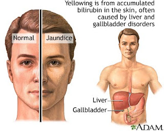
Jaundice , also known as icterus, is a term used to describe a yellowish tinge to the skin and sclerae (the white part of the eye) that is caused by hyperbilirubinemia (an excess of bilirubin in the blood). Body fluids may also be yellow. The color of the skin and sclerae varies depending on levels of bilirubin; mildly elevated levels display yellow skin and sclerae, while highly elevated levels display brown. Bilirubin (bil-ih-ROO-bin) is a yellow colored substance that is responsible for the yellowing of the skin and sclerae. Bilirubin is a waste product that remains in the bloodstream after the iron is removed from the hemoglobin, which is released from the degradation of erythrocytes (cells that contain hemoglobin and can carry oxygen to the body). When there is an excess of bilirubin it may leak out into surrounding tissues, saturating them with this yellow substance. Bilirubin that is circulating freely in the blood is called unconjugated bilirubin. One of the liver's functions is to filter out waste, such as bilirubin, from the blood. Once it is in the liver, other chemicals latch on to the bilirubin, creating a substance called conjugated bilirubin, which is secreted in bile (a digestive juice released by the liver) and then excreted. Bilirubin is what gives feces its brown color. The modern English word "jaundice" is derived from the middle French word jaunisse. Jaun means "yellow" and -isse means "-ness"; hence the middle French word jaunisse, which means "yellowness". According to MediLexicon's medical dictionary, jaundice is: "A yellowish staining of the integument, sclerae, deeper tissues, and excretions with bile pigments, resulting from increased levels in the plasma."
jaundice

There are three main types of jaundice:

Hepatocellular jaundice

- a type of jaundice that occurs as a result of liver disease or injury.

Hemolytic jaundice

- a type of jaundice that occurs as a result of hemolysis (an accelerated breakdown of erythrocytes - red blood cells) leading to an increase in production of bilirubin.

Obstructive jaundice

- a type of jaundice that occurs as a result of an obstruction in the bile duct (a system of tubes that carries bile from the liver to the gallbladder and small intestine), which prevents bilirubin from leaving the liver.
Add Links